
He undergoes the following additional workup for his acute worsening of symptoms:
Labs/Stool studies:
- CRP 18.7 (High), ESR 42.8 (High), WBC 6.7 (normal)
- C-diff: Negative, GI PCR: Negative
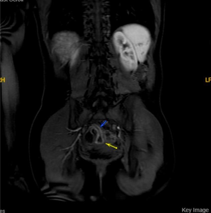
MRE:
- 8 cm of terminal ileal thickening consistent with a known diagnosis of Crohn’s disease. No abscess. No stricture noted.
Colonoscopy:
- Patchy erythema with loss of vascularity throughout the colon, worse in the transverse colon; biopsied.
- Areas of ulceration and edema in the terminal ileum; biopsied. ( Image below)
- Pathology: Active ileitis with ulceration and Chronic, inactive colitis

To move on Click here
